Severe jawbone loss can make conventional implant placement more involved because the tooth-bearing bone may be too thin, too short or too soft for predictable anchorage without additional procedures. By understanding how corticobasal implants are designed, how fixed teeth are delivered and which clinical risks still apply, you can judge whether this approach is worth discussing alongside grafting, sinus lifting or other implant options.
Key takeaways
- Corticobasal implants seek support in denser basal jawbone beneath weakened alveolar bone.
- Fixed teeth are attached to implants and require careful bite, hygiene and maintenance planning.
- A 3D CBCT scan helps assess bone anatomy, nerves, sinuses and implant positioning.
- Severe medical risks, poor hygiene or unsuitable anatomy can make this approach a poor fit.
How corticobasal implants obtain support in weakened jawbone
Conventional implants usually seek primary stability within the tooth-bearing alveolar bone. That bone can become thin, porous and severely resorbed after tooth loss, leaving too little width or height for predictable placement without additional treatment.
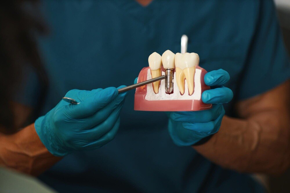
Corticobasal implants are designed to obtain anchorage from denser cortical bone, including the basal cortical bone of the jaw. Basal implants may use longer or laterally positioned fixtures to reach these stronger layers rather than relying only on the weakened central ridge.
| Feature | Conventional implant | Corticobasal implant |
|---|---|---|
| Primary support | Mainly alveolar bone | Dense cortical or basal bone |
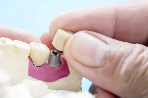
| Design | Usually two-piece, with a separate abutment | Often one-piece, with the abutment integral to the implant |
| Atrophic-jaw strategy | May require added bone or a different implant position | May engage remaining cortical bone when the ridge is severely narrowed |
That distinction can matter in an atrophic jaw because cortical anchorage may provide the mechanical stability needed for a fixed provisional bridge without grafting in a suitable case. Immediate loading is not automatic; it depends on implant stability, bite forces, implant distribution and the planned bridge.
The approach has trade-offs. A one-piece implant offers less opportunity to correct angulation after placement, which can affect bridge design, cleaning access, speech and front-tooth appearance. Three-dimensional imaging must still confirm safe distances from nerves, the sinus and other structures.
What fixed teeth involve after implant placement
After surgery, you may leave the clinic with fixed teeth attached to the implants rather than with removable dentures. This usually means a screw-retained bridge that stays in place during eating and speaking; you cannot take it out for cleaning.
A temporary fixed bridge supports appearance and basic chewing while the tissues settle and the implant-bone relationship is assessed. Immediate loading does not prove that integration has finished. It is safe only when primary stability, implant distribution, splinting, bite control and bridge design limit movement.
| Option | Temporary fixed bridge | Definitive bridge |
|---|---|---|
| Purpose | Early function and protection during healing | Long-term function, appearance and cleanability |
| Timing | Soon after placement when immediate loading is planned | After healing and clinical review |
| Construction | Usually lighter, simpler and easier to modify or replace | More durable, precisely fitted and finished for speech, bite and hygiene |
| Patient removal | Not removed by the patient; the clinician unscrews it | Usually not removed by the patient; the clinician services it |
Treat the provisional bridge as a healing tool, not permission to chew hard foods. Damage, repeated overload or implant movement can jeopardise the result. After healing, the clinician may replace it with a definitive bridge after checking implant stability, bite, speech, cleaning access and appearance.
Ask for the maintenance plan before treatment: daily cleaning beneath the bridge, professional reviews, radiographs when indicated, and adjustment of the bite or prosthetic screws. Fixed teeth are not maintenance-free. One implant problem can affect a connected full-arch bridge.
When this approach may be considered—and when it is a poor fit
Severe bone loss can support consideration when dense cortical bone remains available for stable anchorage, especially around an upper-jaw atrophic ridge or a narrow resorbed ridge in the lower jaw. This approach is not automatically better than bone grafting, sinus lifting, short or tilted conventional implants, or zygomatic implants.
| Situation | Why corticobasal treatment may be considered | What must happen first |
|---|---|---|
| Severe bone loss or a narrow resorbed ridge | Strategic cortical anchorage may support a fixed bridge without rebuilding every deficient area | Confirm adequate bone, implant distribution, bite control and a maintainable prosthetic design |
| Bone grafting or sinus lifting proposed | It may reduce grafting needs when the anatomy allows another stable support pattern | Compare treatment time, cost, surgical risk and the clinician’s experience with alternatives |
| Poor biological conditions | No implant design removes infection or healing risks | Control the underlying problem before surgery |
Smoking, poorly controlled diabetes, untreated periodontal disease and inadequate plaque control require attention before implant placement. A history of severe periodontitis also raises concern because inflammation can affect tissues around any implant, including a polished or smooth-surfaced fixture.
Corticobasal treatment is a poor fit when infection remains active, diabetes is uncontrolled, oral hygiene is inadequate, or the proposed bridge cannot be cleaned and protected from excessive biting forces. Immediate loading also requires primary stability, sound implant distribution, splinting and a controlled occlusion; without them, early movement can jeopardize healing.
A connected fixed bridge adds another consequence: failure of one strategically important implant can affect the bridge segment it supports.
The examination that determines whether the plan is workable
A workable plan begins with three-dimensional anatomy, not with a promise to avoid grafting. The clinician must match the proposed fixtures to available cortical bone, the future bridge and the forces created by your bite.
1. Review a panoramic radiograph for an overview of tooth roots, jaw height, sinus position and obvious pathology. Use a CBCT scan to measure ridge width and height, map dense cortical bone, and identify the inferior alveolar nerve and mental foramen before planning lower-jaw implant positions.
2. Assess the upper jaw’s maxillary sinus and nasal floor. A narrow sinus boundary, an unfavourable nasal floor or insufficient cortical anchorage can change the position, implant design or need for another treatment.
3. Examine your bite, jaw relationship, opposing teeth, smile line, speech needs and available space. These findings determine implant distribution, splinting, bridge contours and whether immediate loading would place damaging movement on healing implants.
4. Check periodontal health, plaque control, smoking, diabetes control, bone infection and your ability to clean beneath a fixed bridge. A graft-free plan does not remove risks such as nerve disturbance, sinus injury, bleeding, infection or damage to adjacent structures.
The clinician should explain why grafting can genuinely be avoided, not simply label the case “atrophic.” Before consent, request the proposed implant positions, loading conditions, provisional and definitive bridge design, alternative treatments, and a maintenance plan covering professional reviews, radiographs when indicated, under-bridge cleaning and bite or screw adjustments.
From consultation to maintenance: the decisions that protect a fixed bridge
Consent should follow a documented examination, not a promise of immediate teeth. Review the CBCT, gum condition, bite, smoking status, medical conditions and cleaning access, then ask how many implants will support the bridge and what happens if one fails.
Before implant placement, agree on:
- Whether primary stability, implant distribution, splinting and bite control justify immediate loading.
- Whether temporary fixed teeth will be fitted the same day or after healing.
- Which parts you can clean or remove, and who removes them.
- The timetable for definitive prosthetic fitting, adjustments and radiographs.
| Plan | Main advantage | Main trade-off and maintenance demand |
|---|---|---|
| Corticobasal bridge | May avoid grafting and provide early fixed teeth in selected atrophic jaws | A connected bridge can be affected by one failing implant; meticulous cleaning and bite reviews remain essential |
| Conventional implants | Larger evidence base and flexible tooth-by-tooth or bridge designs | Severe bone loss may require grafting or sinus lifting, adding healing time and procedures |
| Removable or staged treatment | Easier access for cleaning and repair | Less fixed function, with possible movement and adaptation demands |
Immediate fixed teeth do not prove that osseointegration has finished. Follow the soft-diet and review schedule; biting hard foods, damaging the provisional bridge or overloading an implant can cause mobility.
Ask for written recall intervals, professional cleaning instructions, screw or bite checks and criteria for radiographic monitoring. Plaque can cause peri-implantitis around any implant. Kaira Dental Care & Implant Centre can use this discussion to compare a proposed plan with staged, conventional and removable alternatives rather than treating early loading as the only objective.
Related service
Corticobasal Implants Dr. Ashi Agarwal is a skilled Dental Surgeon offering advanced Corticobasal Implant solutions at her clinic on Wagbil Road, Kavesar, Thane West. View service → |
Frequently asked questions
How do corticobasal implants obtain support in weakened jawbone?
They are designed to engage denser basal or cortical bone rather than relying only on thin, resorbed tooth-bearing alveolar bone.
What do fixed teeth involve after implant placement?
A fixed bridge is attached to the implants, so you cannot remove it like a conventional denture. Its design must support chewing, cleaning and a stable bite.
When may corticobasal implants be considered?
They may be considered when severe jawbone resorption limits conventional implant placement and the examination confirms adequate anatomy and manageable medical risks.
When are corticobasal implants a poor fit?
They are a poor fit when anatomy cannot provide safe support, infection is uncontrolled, hygiene is inadequate, or medical factors make surgery unsafe.
What examination determines whether the plan is workable?
The assessment includes a clinical examination, dental records and three-dimensional CBCT imaging to evaluate bone, nerves, sinuses, infection and the proposed implant path.